Retinal Vein Occlusion Causes, Symptoms, Diagnosis and Treatment

What is Retinal Vein Occlusion?
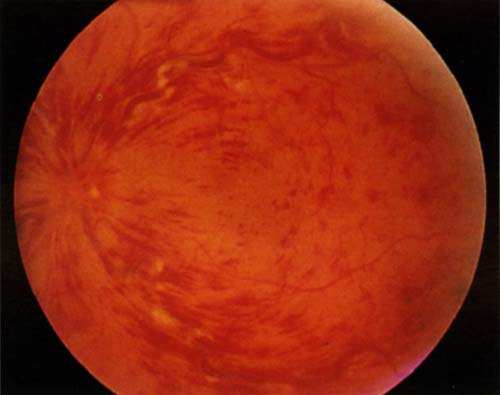
Retinal vein occlusion (RVO) is a partial or total blockage in a vein that drains blood from your retina. Your retina is a layer of tissue at the back of your eye that helps translate light into images you can see. A blockage in a retinal vein prevents blood from leaving your retina. This can lead to complications, including raised pressure in your eye and swelling. These issues need prompt treatment to prevent or minimize vision loss.
There’s no current safe way to unblock the vein. However, treatment can manage complications and protect your vision.
Eye care specialists tailor treatment to your individual needs. You may need multiple retinal vein occlusion treatments ranging from injections to surgery to manage your condition.
Types of retinal vein occlusion
There are two types of RVO:
Central retinal vein occlusion (CRVO), or blockage of the main retinal vein.
Branch retinal vein occlusion (BRVO), or blockage of one of the smaller branch veins. This type is more common.
How common is retinal vein occlusion?
Retinal vein occlusion is the second most common disorder affecting your retina (diabetes-related retinopathy is the most common).
Researchers estimate that globally:
- Retinal vein occlusion affects over 16 million people.
- Central retinal vein occlusion affects between 1 and 4 in 1,000 people.
- Branch retinal vein occlusion affects between 6 and 12 in 1,000 people.
Symptoms of Retinal Vein Occlusion
Symptoms of retinal vein occlusion typically affect one eye and include:
- Blurry vision or vision loss: This may start suddenly or develop gradually over a period of hours or days.
- Floaters: These are dark spots or lines in your field of vision.
- Pain or pressure in your eye: This is typically in more severe cases.
You may not have any symptoms until complications arise. Some people don’t realize there’s a problem until their provider finds the issue during a routine eye exam.
Causes of Retinal Vein Occlusion
A disruption to normal blood flow through your retinal vein causes this condition. The disruption may happen due to:
- A blood clot.
- A slowdown of blood flow.
- Compression of your retinal vein at the point where it crosses paths with your retinal artery. Your retinal artery supplies oxygen-rich blood to your retina. Your retinal artery may grow stiff from aging or plaque buildup, and it may press on your retinal vein. This can damage the inner lining of your retinal vein, creating conditions where a blood clot is more likely to form.
What are the risk factors for retinal vein occlusion?
Being over age 40 is a major risk factor. RVO usually affects people in their 50s or 60s. However, this condition can also affect people younger than age 40.
Having certain medical conditions can also raise your risk. These include:
- Atherosclerosis.
- Diabetes.
- Glaucoma.
- High blood pressure.
Prior history of retinal vein occlusion in one eye raises your risk of developing the condition in your other eye.
What are the complications of retinal vein occlusion?
Retinal vein occlusion can lead to complications such as:
- Cystoid macular edema: This is swelling in the center of your retina (macula). It can cause blurry vision or loss of vision.
- Neovascularization of the eye: Abnormal blood vessels can form in different parts of your eye, typically your iris (rubeosis iridis). This happens in about 1 in 4 people with RVO. Abnormal blood vessels less commonly form in your retina.
- Bleeding in your eye (vitreous hemorrhage): This is when blood leaks into your vitreous humor, the gel-like substance that fills your eyeball. It results from the formation of abnormal blood vessels, which are prone to leaking.
- Neovascular glaucoma: Abnormal blood vessels in your eye can cause pain and a dangerous increase in pressure inside your eye.
- Retinal detachment: Abnormal blood vessels in your retina may cause your retina to pull away from the tissues that support it.
People with RVO have a higher risk of cardiovascular diseases, including stroke, compared to people without RVO. This may be due to shared underlying risk factors like high blood pressure and atherosclerosis.
Diagnosis of Retinal Vein Occlusion
Eye care specialists diagnose RVO through an eye exam and retinal imaging tests. They also coordinate care with your primary care physician (PCP) to discover the cause of blood flow problems.
Eye exam
Your eye care specialist will dilate your pupils so they can see into the back of each eye. They’ll use a microscope and a head-mounted ophthalmoscope to shine a light into your eye. They’ll closely examine the inside of your eye to look for complications and signs of vision loss.
This exam can help:
- Distinguish between central and branch RVO.
- Identify signs of macular edema and abnormal blood vessel formation.
- Estimate how much of your retina lacks blood flow.
You may need further testing to diagnose RVO and show the extent of complications.
Tests to diagnose retinal vein occlusion
Your eye care specialist may use one or more of the following tests to help diagnose and describe your condition:
- Fundus photography: This form of retinal imaging shows the presence of abnormal new blood vessels and the amount of bleeding inside your eye.
- Optical coherence tomography (OCT): This form of high-resolution imaging shows the presence of macular edema. It measures the thickness of your retina and provides precise numbers that help guide the treatment of your condition over time.
- Fluorescein angiography: For this test, your provider injects dye into a vein in your arm. The dye travels to the blood vessels in your retina and makes them stand out in imaging. Your provider may use this form of imaging to show the extent of a blockage in your retinal vein. This test also shows how much of your retina isn’t receiving adequate blood flow.
Coordinated care
Your eye care specialist and primary care physician will work together to find the cause of RVO and lower your risk for future issues. You may need blood tests to check your cholesterol levels, blood sugar and other important numbers.
Treatment of Retinal Vein Occlusion
There’s currently no way to reverse or cure the blockage in your retinal vein. But eye care specialists can prevent or treat the complications of retinal vein occlusion with:
- Anti-VEGF injections.
- Steroid injections.
- Panretinal photocoagulation (PRP).
- Vitrectomy surgery.
- Medications to manage risk factors.
The goals of treatment are to:
- Improve your vision or prevent it from getting worse.
- Identify and treat complications that can harm your vision and eye health.
- Manage risk factors to prevent future problems.
Your provider will combine treatment options as necessary and explain the timing for each.
Anti-VEGF injections
This is a first-line (first-choice) treatment for people with macular edema. VEGF stands for vascular endothelial growth factor. This is a protein that spurs new blood vessel growth (angiogenesis). Too much VEGF can lead to the formation of abnormal blood vessels that can leak and cause swelling.
Anti-VEGF injections interrupt the production of VEGF in your eye to reduce swelling. Your provider gives you eye drops to numb your eye and reduce pain before injecting the medication into the gel-like substance (vitreous humor) that fills your eyeball. You may need injections at regular intervals for one to two years depending on your condition.
Specific medications you may receive in these injections include:
- Aflibercept.
- Bevacizumab.
- Ranibizumab.
Steroid injections
Injections of steroid medication into your eye can also help reduce swelling. However, in some people, steroid injections cause elevated eye pressure and cataracts. So, they’re often a second-line treatment when anti-VEGF injections aren’t adequate.
Panretinal photocoagulation (PRP)
This laser surgery creates small burns in areas of your retina that lack blood flow. Doing so decreases the number of proteins (VEGF) that promote the formation of abnormal blood vessels. Reducing VEGF helps prevent neovascularization and related bleeding in your eye. It also helps keep your intraocular pressure stable.
Vitrectomy surgery
Posterior pars plana vitrectomy (PPV) is a surgery that helps people with retinal vein occlusion who have:
- Severe bleeding in their eye (vitreous hemorrhage).
- Bleeding that lasts more than four weeks.
- Bleeding that keeps coming back.
- Retinal detachment.
Surgery removes vitreous humor from your eye and repairs damage to your retina.
Medications to manage risk factors
Many people with retinal vein occlusion have underlying conditions like high blood pressure, diabetes or high cholesterol. These conditions can raise your risk of blood vessel problems. Your eye care specialist will work together with your primary care physician (PCP) to tailor treatment to your needs. Your PCP may prescribe medications to:
- Lower your blood pressure.
- Manage your cholesterol levels.
- Address other issues.
What can I expect if I have this condition?
Your prognosis depends on many factors, including the location of the blockage and complications that arise. Some people have permanent vision damage, while others have vision that gradually gets better over time. Your eye care specialist is the best person to tell you exactly what you can expect in your individual situation.
Your provider may refer you to vision rehabilitation. This is a form of rehab that teaches you techniques for living with reduced vision. These may include using devices like magnifying glasses or assistive-computer technology. Your provider may also refer you to a social worker who can help you cope with lifestyle changes.
Can I Prevent Retinal Vein Occlusion?
Learning you’re at risk for retinal vein occlusion is the first step toward preventing it. Talk to your ophthalmologist or optometrist about your level of risk and how to lower it.
It’s also important to talk to your primary care physician about underlying conditions that raise your risk for blood flow problems. They’ll recommend treatments as needed to manage those conditions and help keep your eyes — and whole body — healthy.
Specific things you can do to lower your risk include:
- Follow a diet that supports your heart and blood vessel health.
- Make exercise part of your daily routine.
- Keep a weight that’s healthy for you.
- Avoid smoking and all tobacco products.
How do I take care of myself?
Living with retinal vein occlusion (RVO) can be stressful because you may need:
- Multiple eye injections
- Multiple laser treatments.
- Many follow-up appointments.
- Help getting to and from your appointments (if your condition or treatment prevents you from driving safely).
All of this may take a toll and feel overwhelming to you. Remember that your healthcare team is there to help you.
Talk to your providers about how you’re feeling. They may suggest resources to help you learn more about your condition and why all of this effort is so important. They may also connect you with support groups or other community resources where you can talk to people who are in a similar situation. Learning from others’ experiences and sharing your own can help make everything feel more manageable.
When should I seek medical care?
Your eye care specialist will tell you how often you need appointments for monitoring or treatment. Call them if you experience new or changing symptoms or have questions about your treatment plan.
When should I go to the emergency room?
Call 911 or your local emergency number if you have symptoms of a retinal detachment. This is a medical emergency that requires prompt care.
What questions should I ask my provider?
You may want to ask your eye care specialist:
- What caused the blockage in my retinal vein?
- What treatments are best for me?
- What are the benefits and risks of each treatment?
- What follow-ups will I need?
- How will this condition affect my vision?
- What is my outlook?
Reference: https://my.clevelandclinic.org/health/diseases/14206-retinal-vein-occlusion-rvo
By : Natural Health News


